Services

Home Services Brain Services

EPILEPSY SURGERY

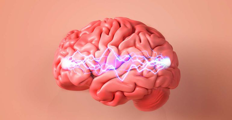
Epilepsy surgery is brain surgery to stop or reduce the number of seizures you’re having and/or their severity. Seizures are a burst of uncontrolled electrical activity between your brain’s nerve cells, which can result in changes in your
   • Awareness
   • Muscle control (your muscles may twitch or jerk)
   • Sensations
   • Emotions
   • Behaviour

Surgical approaches to manage seizures include:
   • Removing the part of your brain where the seizures start
   • Disconnecting brain nerve cell communication to stop the spread of seizures to other areas of your brain
   • Using a laser to heat and kill the nerve cells where the seizures begin
   • Implanting pacemaker-like device & electrodes that send electrical signals disrupting seizure activity at source
   • Inserting delicate electrode wires (using robotic guidance) to record seizure activity from the depths of your brain

A brain aneurysm (AN-yoo-riz-um) — also known as a cerebral aneurysm or intracranial aneurysm — is a bulge or ballooning in a blood vessel in the brain. An aneurysm often looks like a berry hanging on a stem.

Brain aneurysms form and grow because blood flowing through the blood vessel puts pressure on a weak area of the vessel wall. This can increase the size of the brain aneurysm. If the brain aneurysm leaks or ruptures, it causes bleeding in the brain, known as a haemorrhagic stroke.

A brain arteriovenous malformation (AVM) is a tangle of blood vessels that connects arteries and veins in the brain. The arteries take oxygen-rich blood from the heart to the brain. Veins carry the oxygen-depleted blood back to the lungs and heart. A brain AVM disrupts this vital process.

Carotid-cavernous sinus fistula (CCF) is an example of the latter, where a pathologic vascular shunt between the carotid arterial system and the cavernous sinus, or rarely, directly to an ophthalmic or orbital vein. The Barrow classification for CCF divides them into direct (type A) or indirect (types B–D).

A cavernoma is a cluster of abnormal blood vessels, usually found in the brain and spinal cord.

They're sometimes known as cavernous angiomas, cavernous haemangiomas, or cerebral cavernous malformation (CCM). A typical cavernoma looks like a raspberry. It's filled with blood that flows slowly through vessels that are like "caverns". A cavernoma can vary in size from a few millimetres to several centimetres across.

A brain bleed, also known as a brain hemorrhage, refers to bleeding between the brain tissue and the skull or inside the brain tissue. This is a life-threatening condition that requires immediate medical attention. Brain bleeds can limit the oxygen supplied to the brain, causing headaches, nausea, vomiting, tingling in the extremities, or facial paralysis.

A blood clot in the brain stops the flow of oxygen and glucose, causing brain death within minutes. A blood clot in the brain is referred to as a stroke or a brain attack. A stroke happens when the brain's blood vessels burst and bleed, or if the blood flow to the brain is blocked for a few minutes.

MoyaMoya disease is a rare, progressive cerebrovascular disorder caused by blocked arteries at the base of the brain in an area called the basal ganglia. Moyamoya means “puff of smoke” in Japanese and is used to describe the tangled appearance of tiny vessels compensating for the blockage.

The most common type of bypass is the STA-MCA (superficial temporal artery to middle cerebral artery) bypass. The superficial temporal artery (STA) normally provides blood to the face and scalp. You can feel the pulse of the STA in front of your ear. The middle cerebral artery (MCA) normally provides blood to the frontal, temporal and parietal lobes of the brain.

Carotid Endarterectomy (CEA) is surgery to treat carotid artery disease. The carotid arteries are the main blood vessels that carry oxygen and blood to the brain. In carotid artery disease, these arteries become narrowed. This reduces blood flow to the brain and could cause a stroke.

SEEG is the surgical implantation of electrodes into the brain to better localize the seizure focus. Neurosurgeon place several (~10-15) electrodes into your brain. These electrodes are thin, floppy wires about the thickness of a spaghetti noodle. Electrodes are inserted into the brain areas where seizures might be starting. These electrodes are placed with neuronavigation and robotic guidance.

A small incision is made in scalp, followed by drill hole in the bone just big enough to pass the electrode. Each electrode is held in place by a bolt that attaches to the bone. The procedure is done under General anaesthesia. After the procedure patients are in epilepsy monitoring unit. Patients are monitored for couple of days in the hospital and then discharged home after removal of electrodes under mild sedation in operating room.

For more information, please book your appointment with our expert Epileptologist surgeon in Powai Mumbai

Deep brain stimulation (DBS) involves implanting electrodes within areas of the brain. The electrodes produce electrical impulses that affect brain activity to treat certain medical conditions. The electrical impulses also can affect cells and chemicals within the brain that cause medical conditions.

The amount of stimulation in deep brain stimulation is controlled by a pacemaker-like device placed under the skin in the upper chest. A wire that travels under the skin connects this device to the electrodes in the brain.

Deep brain stimulation is commonly used to treat a number of conditions, such as:
• Parkinson's disease
• Epilepsy
• Essential tremor
• Conditions that cause dystonia, such as Meige syndrome
• Obsessive-compulsive disorder
• Tourette syndrome

Deep brain stimulation also is being studied as a potential treatment for:
• Chorea, such as Huntington's disease
• Cluster headache
• Chronic pain
• Dementia
• Addiction
• Refractory depression
• Obesity