CEREBROVASCULAR SURGERY
Cerebro” refers to the large part of your brain and “vascular” refers to your arteries and veins. Cerebrovascular surgery is a specialized surgery performed by a neurosurgeon on the blood vessels that supply blood to your brain.
Sometimes referred to as “neurovascular surgery” and “cerebrovascular neurosurgery,” it’s a treatment for numerous blood vessel conditions and diseases. From addressing restrictions in blood flow to ruptured blood vessels and clots, a cerebrovascular surgeon can perform procedures to help treat vascular conditions on the brain or spine.
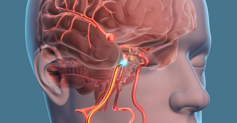
A brain aneurysm also known as a cerebral aneurysm or intracranial aneurysm — is a bulge or ballooning in a blood vessel in the brain. An aneurysm often looks like a berry hanging on a stem, hence they are referred as berry aneurysm.
Brain aneurysms form and grow because blood flowing through the blood vessel puts pressure on a weak area of the vessel wall. This can increase the size of the brain aneurysm. If the brain aneurysm leaks or ruptures, it causes bleeding in the brain, known as subarachnoid haemorrhage (SAH), a kind of haemorrhagic stroke.
This may cause symptoms such as Rapid onset of "worst headache of my life", Stiff neck, Nausea and vomiting, Changes in mental status, such as drowsiness.
A brain aneurysm is often discovered after it has ruptured or by chance during diagnostic exam, such as CT scan, MRI, or angiography that are being done for other reasons.
Brain aneurysms are treated using one or more of the following methods, Microsurgical clipping, endovascular coiling, flow diversion with stents, artery occlusion and bypass surgery.
For more details, please consult our Best neurovascular / cerebrovascular surgeon in Mumbai.
A brain arteriovenous malformation (AVM) is a tangle of blood vessels that connects arteries and veins in the brain. The arteries take oxygen-rich blood from the heart to the brain. Veins carry the oxygen-depleted blood back to the lungs and heart. A brain AVM disrupts this vital process. The most serious complication of DAVF is bleeding in the brain or spine, known as a hemorrhage or hemorrhagic stroke. The most serious complication of AVM is bleeding in the brain or spine, known as a hemorrhage or hemorrhagic stroke.
AVM are treated using one or more of the following methods, Microsurgical excision, endovascular coiling or embolization and or Stereotactic radiotherapy.
For more details, please consult our Best Brain and spine specialist doctor in Powai, Mumbai.
AVM are treated using one or more of the following methods, Microsurgical excision, endovascular coiling or embolization and or Stereotactic radiotherapy.
For more details, please consult our Best Brain and spine specialist doctor in Powai, Mumbai.
A dural arteriovenous fistula (DAVF) are abnormal connections between arteries and veins in the membrane (Dura) that covers the brain and spinal cord.
Dural AV fistulas are a type of arteriovenous malformation (AVM). An arteriovenous malformation, or fistula, is direct connection between an artery and a vein. Usually, a bed of capillaries separates arteries and veins. A DAVF bypasses the capillaries, which can lead to high pressure in the receiving veins.
The most serious complication of DAVF is bleeding in the brain or spine, known as a hemorrhage or hemorrhagic stroke.
Your treatment will depend on your symptoms, overall health and risk of hemorrhage. If you have a low grade, benign DAVF, your doctor may opt to observe you over time. You may never need treatment.
Higher-risk DAVFs usually require surgical treatment to disconnect the arteriovenous connection. Our neurosurgeons offer the most advanced treatments available today, including: endovascular embolization, microsurgery, radiosurgery.
For more details please book your appointment with our Best Brain and spine specialist in Thane and Mumbai.
A cavernoma is a cluster of abnormal blood vessels, usually found in the brain and spinal cord.
They're sometimes known as cavernous angiomas, cavernous haemangiomas, or cerebral cavernous malformation (CCM). A typical cavernoma looks like a raspberry. It's filled with blood that flows slowly through vessels that are like "caverns". A cavernoma can vary in size from a few millimetres to several centimetres across.
A cavernoma often does not cause symptoms, but when symptoms do occur they can include:
• bleeding (haemorrhagic stroke)
• fits (seizures)
• headaches
• neurological problems, such as dizziness, slurred speech (dysarthria), double vision, balance problems and tremor
• weakness, numbness, tiredness, memory problems and difficulty concentrating
Cavernomas are diagnosed using MRI. Treatment for cavernomas includes:
• Medication to treat symptoms like seizure or headache.
• Surgery remove your cavernoma if you're experiencing symptoms.
• Genetic testing and counselling is advisable in familial cases of cavernoma.
For more details, please book your appointment with our Best neurosurgeon in Thane and Mumbai.
A brain bleed, also known as a brain hemorrhage, refers to bleeding between the brain tissue and the skull or inside the brain tissue. This is a life-threatening condition that requires immediate medical attention. Brain bleeds can limit the oxygen supplied to the brain, causing headaches, nausea, vomiting, tingling in the extremities, or facial paralysis.
Types of Brain Bleeds
Bleeding within the skull but outside the brain tissue
• Epidural hemorrhage -
• Subdural hemorrhage -
• Subarachnoid hemorrhage
Bleeding inside the brain tissue
• Intracerebral hemorrhage - Bleeding that occurs in the cerebellum of the brain (including the brainstem)
• Intraventricular hemorrhage - Bleeds that originate in the brain cavities where cerebrospinal fluid is produced.
The symptoms of a brain hemorrhage can vary. They depend on the location of the bleeding, the severity of the bleeding, and the amount of tissue affected. Symptoms tend to develop suddenly.
Treatment for bleeding in the brain depends on the location, cause, and extent of the hemorrhage. Surgery may be needed to alleviate swelling and prevent bleeding. Certain medications may also be prescribed. These include painkillers, corticosteroids, or osmotics to reduce swelling, and anticonvulsants to control seizures.
For more information please book your appointment with our Consultant neurosurgeon in Powai Mumbai.
MoyaMoya disease is a rare, progressive cerebrovascular disorder caused by blocked arteries at the base of the brain in an area called the "Circle of Willis". Moyamoya means “puff of smoke” in Japanese and is used to describe the tangled appearance of tiny vessels compensating for the blockage.
People with moyamoya disease presents with ischemic stroke, hemorrhagic stroke, and seizures.
In diagnosing moyamoya disease, the doctor may recommend an EEG, CT, MRI of the brain or Cerebral angiography to confirm the diagnosis.
Moyamoya disease treatment involves managing symptoms, improving blood flow to the brain and controlling seizures.
In some cases, Neurosurgeon perform revascularization surgery to rebuild the blood supply to the underside of the brain, a delicate and technically demanding procedure that should be performed by an expert neurosurgeon.
For more information please book your appointment with our expert neurosurgeon in Powai Mumbai.
The most common type of bypass is the STA-MCA (superficial temporal artery to middle cerebral artery) bypass. The superficial temporal artery (STA) normally provides blood to the face and scalp. You can feel the pulse of the STA in front of your ear. The middle cerebral artery (MCA) normally provides blood to the frontal, temporal and parietal lobes of the brain.
When blood circulation in the brain is compromised due to blockage or narrowing of the carotid artery or it's branches, STA is anastomosed with MCA branch to augment intracranial circulation. This is similar to bypass surgery done on heart for coronary artery disease.
For more information please book your appointment with our expert Cerebrovascular neurosurgeon in Mumbai.
Carotid Endarterectomy (CEA) is surgery to treat carotid artery disease. The carotid arteries are the main blood vessels that carry oxygen and blood to the brain. In carotid artery disease, these arteries become narrowed. This reduces blood flow to the brain and could cause a stroke.
By means of CEA blocked carotid arteries are reopened by removing the plaques in the lumen of the diseased artery.
For more information please book your appointment with our expert Cerebrovascular neurosurgeon in Mumbai.
By means of CEA blocked carotid arteries are reopened by removing the plaques in the lumen of the diseased artery.
For more information please book your appointment with our expert Cerebrovascular neurosurgeon in Mumbai.